
Globally, Trichuris trichiura, or whipworm, is a very common intestinal helminthic infection, and about one quarter of the world's population is thought to carry the parasite. Principally a problem in tropical Asia and, to a lesser degree, in Africa and South America, a lack of a tissue migration phase and a relative lack of symptoms characterize whipworm infection. Trichuris is also notable for its small size compared with Ascaris lumbricoides. Only patients with heavy parasite burden become symptomatic. Vitamin A deficiency has been seen in patients with T trichiura infection .
Poor hygiene is associated with trichuriasis transmission, and children are especially vulnerable because of their high exposure risk. This is especially true in developing countries, where poor sanitary conditions correlate with heavy disease burden and infections. One study in Nigeria was undertaken to determine helminth infection status and hygienic conditions in primary schools. Prevalence of helminth infection was higher in the schools where hygiene conditions (ie, tapwater, handwashing soap) were lacking. The study results recommended that the school health programs include deworming, health education, and improvement of hygiene conditions.[1]
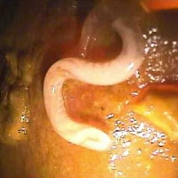
The whipworm derives its name from its characteristic whiplike shape; the adult (male, 30-45 mm; female, 35-50 mm) buries its thin, threadlike anterior half into the intestinal mucosa and feeds on tissue secretions, not blood. This relative tissue invasion causes occasional peripheral eosinophilia. The cecum and colon are the most commonly infected sites, although in heavily infected individuals, infection can be present in more distal segments of the GI tract, such as the descending colon and rectum. See the image below.
Poor hygiene is associated with trichuriasis transmission, and children are especially vulnerable because of their high exposure risk. This is especially true in developing countries, where poor sanitary conditions correlate with heavy disease burden and infections. One study in Nigeria was undertaken to determine helminth infection status and hygienic conditions in primary schools. Prevalence of helminth infection was higher in the schools where hygiene conditions (ie, tapwater, handwashing soap) were lacking. The study results recommended that the school health programs include deworming, health education, and improvement of hygiene conditions.[1]
The whipworm derives its name from its characteristic whiplike shape; the adult (male, 30-45 mm; female, 35-50 mm) buries its thin, threadlike anterior half into the intestinal mucosa and feeds on tissue secretions, not blood. This relative tissue invasion causes occasional peripheral eosinophilia. The cecum and colon are the most commonly infected sites, although in heavily infected individuals, infection can be present in more distal segments of the GI tract, such as the descending colon and rectum. See the image below.
Adult males of Trichuris trichiura are 30-45 mm long, with a coiled posterior end. Adult females are 35-50 mm with a straight posterior end. Both sexes have a long, whip-like anterior end. Adults reside in the large intestine, cecum, and appendix of the host. Image shows the posterior end of an adult T trichiura, taken during a colonoscopy. Image courtesy of Duke University Medical Center and Centers for Disease Control and Prevention.


0 komentar:
Posting Komentar